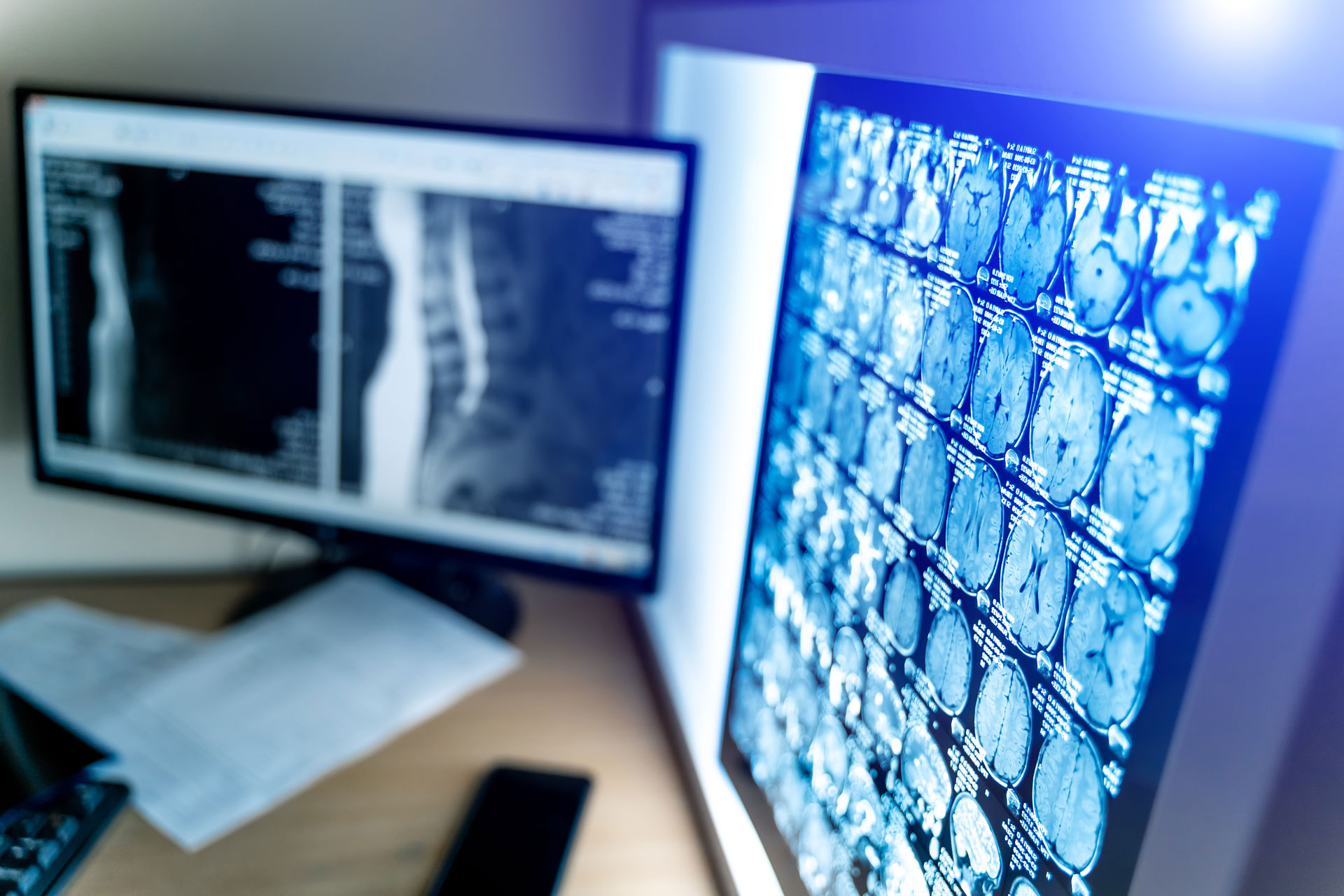
Medical imaging, particularly Magnetic Resonance Imaging (MRI), has revolutionized diagnostic medicine, providing detailed insights into the human body. Among the various phenomena observed in these images, T2 hyperintensity stands out as a critical indicator. Therefore, understanding what t2 hyperintensity mean with the causes of T2 hyperintensity is paramount for accurate diagnosis and effective treatment planning.
T2 Hyperintensity Unveiled
T2 hyperintensity refers to brighter signals on T2-weighted MRI images. It often signifies abnormalities in the underlying tissues, allowing healthcare professionals to pinpoint potential issues. Let’s delve into the primary causes of T2 hyperintensity and how it manifests in different MRI settings.
1. Inflammation and Edema
Primarily, inflammatory conditions and edema are frequent culprits behind T2 hyperintensity. As a result of increased fluid content, characteristic of inflammation and edema, there is a heightened signal intensity on T2-weighted images. This is commonly observed in diseases like arthritis, tendonitis, and infections affecting joints or soft tissues.
2. Vascular Abnormalities
Secondly, malformations in blood vessels can induce T2 hyperintensity. Conditions like arteriovenous malformations (AVMs) and aneurysms alter blood flow, leading to changes in tissue characteristics visible on MRI hyperintensity scans. Therefore, identifying such abnormalities is crucial for planning appropriate interventions.
3. Neurological Disorders
Thirdly, certain neurological disorders exhibit T2 hyperintensity in the brain or spinal cord. Conditions like multiple sclerosis (MS), cerebral infarction, and gliomas often manifest as hyperintense regions on T2-weighted images. Analyzing the location and pattern aids in diagnosing and managing these disorders effectively.
4. Ischemia and Infarction
Fourth, insufficient blood supply, as seen in ischemic conditions, can result in T2 hyperintensity. Areas with compromised blood flow, leading to tissue damage, appear brighter on T2-weighted images. Recognizing these patterns is crucial for understanding the extent of damage and guiding therapeutic decisions.
5. Neoplastic Growth
Last, tumors, both benign and malignant, frequently display T2 hyperintensity. The increased water content in tumors contributes to the heightened signal intensity. Also, analyzing the location, size, and morphology aids in differentiating between various types of neoplasms.
Open MRI and T2 Hyperintensity
Furthermore, the choice of MRI equipment can influence the visibility and interpretation of T2 hyperintensity. Open MRI machines, characterized by a more spacious design, are often preferred for patients with claustrophobia or larger body types. However, the open configuration may impact the image quality, potentially affecting the detection of T2 hyperintensity.
In an open MRI, the surrounding air may lead to increased susceptibility artifacts, causing signal variations that can be misinterpreted as T2 hyperintensity. Therefore, radiologists need to be aware of these potential artifacts and exercise caution when interpreting open MRI images for T2 hyperintensity-related findings.
Wide-Bore MRI: A Closer Look
Wide-bore MRI machines, designed to accommodate larger patients, offer a compromise between open and traditional closed MRI scanners. While they provide a more spacious environment, akin to open MRI, they also minimize some of the challenges associated with image quality.
In wide-bore MRI, the impact of the surrounding air is reduced, mitigating susceptibility artifacts. This can enhance the accuracy of T2-weighted images, aiding in a more precise diagnosis of T2 hyperintensity-related conditions. Radiologists may find wide-bore MRI particularly advantageous when scrutinizing areas prone to artifacts, such as the joints.
Choosing the Right MRI for T2 Hyperintensity Evaluation
Now, let’s talk about selecting the appropriate MRI machine for assessing T2 hyperintensity. Well, it depends on various factors, including patient comfort, the area of interest, and the clinical context. While open MRI provides a more comfortable experience for certain patients, radiologists must be vigilant regarding potential artifacts that might complicate T2 hyperintensity interpretation.
Wide-bore MRI offers a balanced solution, catering to patient comfort while maintaining image quality. Healthcare providers need to weigh these factors and choose the MRI technology that best suits the specific diagnostic requirements.
Innovations in Imaging: The Future of T2 Hyperintensity Analysis
As technology advances, so does the field of medical imaging. Innovations in MRI technology continue to refine the accuracy and efficiency of T2 hyperintensity analysis. One promising avenue is the integration of artificial intelligence (AI) and machine learning algorithms into image interpretation.
AI algorithms can assist radiologists in identifying subtle patterns associated with T2 hyperintensity, potentially reducing the risk of oversight or misinterpretation. Further, these tools can learn from vast datasets, enhancing their ability to recognize nuanced variations indicative of different medical conditions.
Moreover, real-time processing and reconstruction techniques are evolving, allowing for quicker and more detailed image acquisition. This is particularly relevant in situations where timely diagnosis is critical, such as acute neurological events or emergent vascular abnormalities.
Patient-Centric Care: Navigating T2 Hyperintensity Anxiety
For patients undergoing MRI examinations, concerns about T2 hyperintensity findings can lead to anxiety. Understanding the nature of T2 hyperintensity and its various causes is essential in alleviating these concerns. Healthcare providers play a crucial role in educating patients about the significance of these findings and the subsequent steps in the diagnostic and treatment process.
The choice between open MRI, wide-bore MRI, or traditional closed MRI may also factor into addressing patient anxiety. New Jersey Imaging Network Edison recognizes the importance of patient-centric care, offering a range of imaging options tailored to individual needs. The commitment to providing a comfortable and reassuring environment is integral to fostering trust between patients and healthcare providers.
Challenges in Interpretation and Standardization
While T2 hyperintensity is a valuable diagnostic tool, challenges persist in its interpretation and standardization. For instance, variability in imaging protocols, equipment specifications, and individual patient factors can influence the appearance of T2 hyperintensity. Moreover, standardizing imaging techniques and developing comprehensive guidelines are ongoing challenges in the field.
Hence, radiologists must remain vigilant in distinguishing true pathology from artifacts or benign variations in T2 hyperintensity. Collaborative efforts within the medical imaging community are essential to establish consensus guidelines that enhance the reliability and reproducibility of T2-weighted images across different imaging platforms.
Clinical Correlations: Beyond T2 Hyperintensity
In clinical practice, T2 hyperintensity is rarely considered in isolation. Radiologists analyze T2-weighted images in conjunction with other imaging sequences, clinical history, and laboratory findings to form a comprehensive diagnostic picture. The integration of multi-parametric imaging approaches enhances the accuracy of diagnoses and facilitates a more holistic understanding of the patient’s condition.
Moreover, longitudinal studies are shedding light on the dynamic nature of T2 hyperintensity. Tracking changes in hyperintense regions over time provides valuable information about disease progression and response to treatment. This temporal dimension adds a layer of complexity to the interpretation of T2-weighted images, necessitating a nuanced and patient-specific approach.
Conclusion:
In the realm of medical imaging, understanding the causes of T2 hyperintensity is crucial for accurate diagnosis and treatment planning. Whether utilizing open MRI, wide-bore MRI, or traditional closed MRI for T2 hyperintensity, each technology has its nuances in detecting and interpreting T2 hyperintensity.
For residents in New Jersey seeking affordable imaging solutions, the New Jersey Edison Imaging Network stands as a reliable resource for open MRI in New Jersey. Committed to affordability and excellence, NJ Imaging Center employs cutting-edge technology to ensure accurate diagnoses while prioritizing patient comfort.
In the dynamic landscape of medical imaging, staying informed about T2 hyperintensity and the technologies involved is paramount. The journey through the intricate details of T2 hyperintensity continues, shaping the way healthcare professionals approach diagnosis and patient care.

