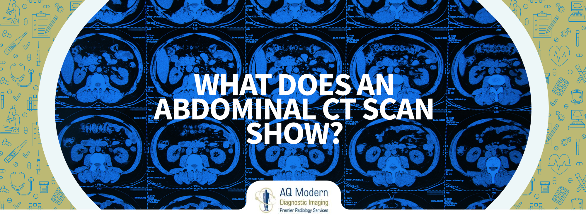
Abdominal CT scan
A CT scan, sometimes referred to as a CAT scan or a CT scan, is a kind of diagnostic imaging network. It is one of the most affordable imaging techniques. The pictures produced by a CT scan may be re-formatted in a variety of ways, and a three-dimensional view may also be generated. Images may be seen on a computer display, printed on film or 3D printed, or burned to a CD or DVD for your doctor’s inspection.
CT scans of bones, internal organs, soft tissue, and blood vessels are superior to standard x-rays in terms of detail.
In What Situations Is Abdominal CT Scan Most Often Used?
Abdominal and pelvic discomfort is often treated using CT scan imaging by doctors. The small intestine and colon, as well as appendicitis, pyelonephritis, and infected fluid collections, known as abscesses, may also be diagnosed using CT scan diagnostic services.
Pancreatitis, ulcerative colitis, cirrhosis of the liver, and other inflammatory bowel diseases are also detected using CT scan services.
In addition to lymphoma, abdominal CT scan detect:
- Malignancies of the ovaries and bladder.
- Urinary tract and kidney stones.
- Injury to the abdomen, such as liver, spleen, or kidneys, may develop an abdominal-abdominal aortic aneurysm.
Your doctor also prescribes abdominal CT scans to:
- Design and evaluate surgical outcomes, such as organ transplantation.
- Guides biopsies and other operations such as abscess drainages and less invasive tumor therapies.
- Staged, planned, and correctly administered Tumor radiation treatments, and the effectiveness of chemotherapy is monitored as a result.
How To Prepare For An Abdominal CT Scan?
Wear a loose-fitting, comfortable dress for the abdominal CT scan. CT scans for abdominal pain may be distorted if they contact metal items, such as jewelry, eyeglasses, dentures, or hairpins. They should either be left at home or removed before the test. Some abdominal CT scan examinations need the removal of removable dental and hearing aids.
If your abdominal CT scan includes the use of contrast material, your doctor may advise you to refrain from eating or drinking for another few hours beforehand. Make sure your doctor is aware of all the medicines you are currently taking and any allergies you may have. Your doctor may prescribe prescription drugs if you’ve been diagnosed with an adverse response to contrast material. Before your checkup, call your doctor to minimize any needless delays.
Be sure to let your doctor know if you’ve recently been unwell or have any other medical concerns such as heart or thyroid problems or a history of diabetes, renal disease, or asthma. Any of these factors might enhance the chance of an undesirable impact.
Pregnant women should always tell their doctor and the abdominal CT scan technician.
What Is The CT Scanner Like?
The abdominal CT scan machine is like a large donut-shaped machine with a small tube in the middle. You’ll be strapped to a table that glides in and out of a tight passageway. The x-ray tube and electronic x-ray detectors are arranged in a gantry that revolves around you. There is a separate control room for the computer workstations that process the image information. This is where the technician runs the scanner and watches your exam in real-time. In order to communicate with you, the techie will use a speaker and microphone.
How Does The Procedure Work?
A Full Body CT Scan NJ is a lot like other x-ray tests, and various body regions absorb x-rays at varying rates. Because of this disparity, a CT picture may be used to differentiate different bodily sections from one another.
Radiation from a normal x-ray exam is focused on the bodily portion being examined. The picture is captured using a specialized electronic image recording plate. X-rays show white bones. Soft tissue, such as the heart and liver, appears as a grayscale image, and the air looks black.
During CT scan services in NJ, you are surrounded by a rotating array of x-ray beams and electronic x-ray detectors. The quantity of radiation absorbed by your body is determined by using one of these devices. Occasionally, the scanning table may shift. This massive amount of data is processed by specialized computer software to produce cross-sectional photographs of your body in two dimensions. The photographs are shown on a computer monitor by the system. Computer software reassembles picture slices to provide a multidimensional depiction of the body’s inside that is very accurate and precise.
Almost all CT scanners can acquire numerous slices within a single spin. These CT scanners with several slices may produce thinner slices in less time.
Large parts of the body may be imaged in only a few seconds by modern CT scanners and even quicker in little children. All patients benefit from this level of efficiency. Speed is particularly advantageous for youngsters, the elderly, and the seriously sick – anybody who is unable to remain motionless for the small period of time required to capture photographs.
Using a smaller CT scanner, the radiologist will be able to limit radiation exposure for youngsters. Occasionally, a contrast substance is injected into the region being examined during an abdominal CT scan.
How CT Scan is Performed?
The technician starts by adjusting your position on the CT exam table, which is typically flat on your back. They may employ straps and cushions to keep you in the ideal posture when doing an exam.
Many scanners are quick enough to do so when scanning children without anesthesia. The use of sedatives may be necessary for kids who cannot remain motionless, and the motion may generate blurry images and reduce the image quality as with pictures.
In certain cases, the test may employ contrasting materials. If this is the case, it will be given orally, intravenously (IV), or by enema. Then, the table will be scanned rapidly to identify where to begin the scans. The CT scanner will move the table gently through the machine, and the scanner may make numerous passes, depending on the kind of CT scan used.
You may be asked to hold your breath by the technician during the scanning process. Any movement, including breathing or body motions, might cause artifacts in the photographs. This loss in picture quality might be likened to the blurring while photographing a moving object.
When the test is completed, the technician will ask you to wait until the pictures are of sufficient quality for accurate description by the radiologist.
The CT scan normally just takes a few minutes to complete. The imaging facility will ask you to come around two hours before your scan appointment if you are required to consume an oral contrast solution before your scan.
What To Expect During And After The Surgery?
CT scan NJ is typically painless and quick. Although the scan is harmless, holding motionless for many minutes or placing an IV may cause discomfort. A CT test might be uncomfortable if you have trouble sitting still, are apprehensive, anxious, or in pain. A technician or nurse may prescribe you medicine to assist you in enduring the CT exam.
In this case, your doctor will look for chronic renal illness. The doctor gives intravenous contrast material and causes a pinprick when injected. The contrast may cause a warm or flushed feeling, and you may also taste metallic.
You may find it uncomfortable to take oral contrast material. But most patients can tolerate it. An enema will cause abdominal fullness. You may start to feel the need to vomit more often. Be patient; the slight pain will pass.
Many patients also get an intravenous iodine-based contrast material to examine blood arteries and organs such as the liver, kidneys, and pancreas.
The CT scanner may produce specific light lines over your body. These lines help you get on the test table correctly. Modern CT scanners make a buzzing, clicking, and whirring noise. During the imaging procedure, interior pieces of the CT scanner whirl around you. You will be the only patient in the CT scan room unless an exception is made. Some parents wear lead shields and remain in the room with their kids. The technician can always see, hear, and communicate with you through an intercom system.
The technician will remove your IV after the CT scan, and they will treat the needle’s tiny hole. You may resume routine activities right away.
What Happened After The Scan?
An expert will examine images in the field of radiology. The doctor who requested the test will get an official report from the radiologist.
It is possible that you may require a follow-up examination. If this is the case, your physician will explain why. A follow-up test provides further information about a suspected problem in certain cases. It may also check to determine whether a problem has changed over time—the easiest approach to determine whether therapy is effective or the problem is to schedule follow-up tests.

