
Have you ever wondered what you’re looking at when looking at an ultrasound image? Have you ever wondered which way is up? Are you still trying to figure out what it is when others are talking about bull or heifer, ovarian diagnostic, or metritis treatment? So, if you’re feeling a little lost in ultrasound photos, let’s start from the beginning.
Many people find it difficult to understand ultrasound interpretations. Knowing how to interpret the results, on the other hand, can help you educate both other staff members and patients. Ultrasound images reading is not that challenging if you have the basic knowledge. The radiologists and doctors are provided with sufficient training on how to read an ultrasound.
Medical imaging has aided in transforming lives and the prevention of potentially fatal illnesses. Ultrasound is used to diagnose and prevent illnesses, but it can also be used for other therapeutic purposes, which will be discussed further below.
Ultrasound Images Reading
Did you recently have an ultrasound? Want to know how to read an ultrasound? We know that ultrasound images look like a puzzle for common people without medical knowledge. You might be trying to interpret it, but it requires some understanding. For the untrained eyes, it’s more challenging.
An ultrasound may be performed for various reasons, the most common of which is to view a baby in the womb. You may also be interested in learning how to identify specific features of your pregnancy ultrasound, such as the baby’s head, arms, or sex.
Once the ultrasound is performed, we know people try to figure out how to read ultrasound reports. They focus on finding ways how to read ultrasound images. It’s not very complicated but requires attention and understanding.
Here are some tips that can help in ultrasound image reading:
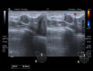
1. Ignore the Text and Numbers on the Top
The hospitals use the numbers and text on the top for reference. It might include the details of ultrasound machine settings. It does not have anything to do with you.
2. Begin from the Top
Now, you look at the top of the image. It shows where the probe was placed during the ultrasound. Here you can see the organs or tissues.
For instance, at the top of the ultrasound images of your uterus, you’ll see the outline of tissues present in the uterus. However, if you go a bit down the screen, you’ll find the deeper tissue and lining of the uterus.
3. Focus on the Colors and their Differences
Most ultrasound images are black and white, but the shades of black and white in your ultrasound scan can vary. The color differences are caused by differences in the densities of the materials through which the sound travels.
During ultrasound images reading, you’ll find the following:
- Solid tissues, including the bones, appear white.
- The tissues have liquid, so they appear dark.
4. Find Out the Visible Side of the Body
During ultrasound images reading, you must know that these are imaged. The left side of the body is on the left side of the image.
5. Focus on the Visual Effects
The ultrasound technique is used for assessing the inner structures of the body. It uses sound to create images. However, these images aren’t very clear. You’ll find various visual effects that rely on the radiologist or doctor’s settings, angles, and density set.
Here are a few things that you can look for:
- Attenuation – it’s the shadowing effect that makes the scanned area appear darker.
- Enhancement – through this, the examined area looks brighter.
- Anisotropy – relies on the angle of the probe. The doctors should adjust the right angle.
6. Assessing the Structures
To interpret structures, you’ll require some knowledge of human anatomy. For instance, if you want to determine the sex of the baby in the womb, you’ll have to focus on some specific shapes and structures.
The sonographers are experienced in identifying the structures. You might need their help with the assessment.
Ultrasound Picture Explained
We’ve included more information if you’re still confused about ultrasound image reading. Go on reading and find the ultrasound picture explained in more detail:
Firstly, the baby will look grey or white in the image. It’s because the baby is located in amniotic fluid.
- The 8-week ultrasound shows the size of the fetus, similar to a baked bean.
- In the 12-weeks ultrasound, you’ll see the head of the baby.
- In 20-weeks, you can see the baby’s heart, feet, eyes, and spine.
Overall, the sonogram technicians are capable of revealing the baby’s gender. From the ultrasound picture explained, it becomes easier to assess and monitor the growth and development of the fetus. You can consider opting for a 3D or 4D ultrasound scan for better results and more details.
When focusing on ultrasound images reading, you’ll see the following information:
- Gestational age of the fetus.
- The length between both sides of the head.
- Measurements of the femur and thighbone.
- Height and weight of the fetus.
- Fetal measurements to assess any abnormalities in the structure.
Final Thoughts
The bottom line is that ultrasounds are the first things that come to mind during pregnancy. However, your doctor might suggest an ultrasound for other reasons too. It also helps in diagnosing and preventing illnesses like other imaging techniques.
Once you’re done with an ultrasound, we know most to-be-mothers try to learn ultrasound images reading. They use the internet to learn how to read ultrasound reports. For this, we have summed up some important information that can help.