For anyone dealing with knee pain due to any injury or condition can be painful and discerning. In most cases, doctors recommend getting a knee MRI (Magnetic Resonance Imaging) to diagnose the root cause of the problem. An MRI is a non-invasive, secure imaging method that offers detailed pictures of the knee joint and the tissues around it.
In this comprehensive article, we will delve into various aspects of knee MRI, covering essential information such as the procedure itself, MRI time length, necessary preparation beforehand, and what you can anticipate during the examination process.
Understanding the Knee MRI Procedure
What is a Knee MRI?
A Knee MRI is simply an imaging procedure of the knee to generate images that a physician can analyze to diagnose the cause of the problem. It relies on powerful magnetic and radio waves to produce comprehensive visuals of the joints.
The MRI of the knee does not use ionizing radiation as is used in procedures like CT scans or X-rays. Therefore, it is safe and preferable for evaluating soft tissues and internal structures.
Why is a Knee MRI Performed?
The MRI machine takes extensive images of your knee and the surrounding area of your muscles, joints, and tissues, which helps the healthcare provider understand the problem deeply.
Your physician may order a knee MRI to rule out various conditions, as knee pain can result from various factors. Typically, a knee MRI will be done in case of the following issues:
- Ligament or tendon tears
- Meniscal injuries
- Cartilage abnormalities
- Bone fractures
- Arthritis
- Joint infections
- Tumors or cysts
- Sports-related injuries
- Fluid buildup
- Infection
- Problems with medical device implants
By obtaining clear and precise images, a knee MRI helps healthcare professionals accurately diagnose these conditions and devise appropriate treatment plans.
Preparing for a Knee MRI
Before your knee MRI, following the preparation guidelines provided by your healthcare provider is essential. These instructions may include:
- Informing the technician about any metal implants or medical devices you have, such as pacemakers, artificial heart valves, or cochlear implants.
- Wearing comfortable clothing without metal zippers, buttons, or jewelry.
- Removing any metallic objects, such as watches, belts, and keys.
- Talk about any allergies or additional issues you may have, such as claustrophobia.
- If necessary, refrain from food and liquids for a predetermined amount of time before the treatment.
- Following these guidelines ensures a smooth and accurate MRI examination.
The Duration of a Knee MRI
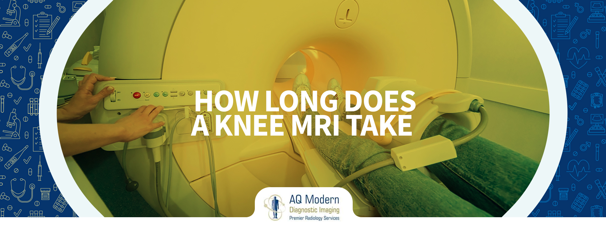
One of the common concerns individuals have before undergoing a knee MRI is the duration of the procedure. On average, a knee MRI takes approximately 30 to 45 minutes. However, the exact duration may vary depending on several factors:
- The complexity of the knee condition that is being evaluated.
- The number of images required.
- The patient’s capacity for maintaining stillness during the scan.
- The imaging facility’s particular protocols and methods.
It’s important to note that additional imaging sequences or contrast agents may be required in some cases. This can extend the examination time.
What to Expect During a Knee MRI
When you arrive at the imaging facility for your knee MRI, a radiology technician will greet you and guide you through the process. Here’s what you can expect during the procedure:
- In order to prevent metal items from interfering with the MRI, you will likely be required to change into a hospital gown.
- An imaging coil or other specific gadget will be put around your knee while you are lying on a mobile examination table.
- The technician will appropriately position the table and coil and ensure your comfort throughout the procedure.
- Once you are ready, the table will slide into the MRI scanner, which is a large tube-shaped machine.
- In order to get clear images from the scan, it’s crucial to maintain as much stillness as possible. Some sequences may need you to temporarily hold your breath.
- Throughout the test, the MRI machine makes a series of loud pounding or thumping noises. To reduce noise, earplugs or headphones will likely be offered.
- If needed, the technician will communicate with you through an intercom system to provide instructions or check your well-being.
- Once the scan is complete, the table will slide out of the MRI machine, and the technician will assist you in getting up.
What Does A Knee MRI Feel Like?
When you are inside the MRI machine, you will not really feel it working. However, you may feel a cooling sensation after applying the contrast dye. There are no other physical feelings that you may feel.
As the machine starts, you may experience noises like humming, thudding, or whirring. If you are at a reputable Diagnostic Imaging Network, your technician may give you headphones to help diminish the sound allowing you to feel more comfortable during the procedure.
Risks of MRI: Understanding the Potential Concerns
Detailed pictures of the body’s interior structure can be acquired through the commonly used diagnostic imaging technology known as magnetic resonance imaging. It is considered safe for most individuals. However, like any medical procedure, MRI has potential risks and considerations, as follows:
MRI and Claustrophobia
One of the most common concerns associated with MRI is claustrophobia. The MRI machine has a narrow tunnel-like structure, which can cause anxiety and discomfort for individuals who experience claustrophobic tendencies. Panic attacks or increased anxiety can be brought on by the sense of confinement in a small area.
To alleviate this concern, many imaging facilities offer Open MRI Nj scanners that are more spacious and less restrictive. These scanners provide a more comfortable experience for individuals prone to claustrophobia. It is essential to discuss your claustrophobia concerns with your healthcare provider or imaging facility beforehand, as they can guide you toward the most suitable imaging option.
Potential Allergic Reactions
Another potential risk associated with MRI involves the use of contrast agents. Contrast agents are chemicals administered intravenously to the body during a scan to make specific tissues or blood vessels more visible. They help provide a clearer image and assist in diagnosing specific conditions.
Although rare, some individuals may have an allergic reaction to contrast agents. It is essential to let your healthcare professional know if you have any known allergies before having an MRI, especially if the allergy is to contrast materials. With the use of this knowledge, the medical staff is able to take the proper safety measures and, if required, select other imaging techniques.
Metal Implants and MRI Safety
MRI machines utilize strong magnetic fields, which can interact with metallic objects within the body. It is vital to inform your healthcare provider and the MRI technologist about any metal implants, devices, or objects in your body, as they may pose risks during the scan.
Common metal implants that may interfere with an MRI include:
- Pacemakers or implantable cardioverter-defibrillators (ICDs)
- Cochlear implants
- Artificial heart valves
- Joint replacements (e.g., hip or knee implants)
- Surgical clips or wires
These metal objects can potentially malfunction or shift due to the magnetic field, leading to serious complications. In some cases, individuals with such implants may not be eligible for an MRI. Communicating openly with your healthcare provider to determine the most suitable imaging method based on your medical history and implanted devices is essential.
Pregnancy and MRI
MRI scans are generally safe to conduct during pregnancy, particularly in the second or third trimester. However, as a precautionary measure, it is advisable to avoid MRI typically during the first trimester unless it is absolutely necessary.
While no harmful effects of MRI on the developing fetus have been established, it is crucial to inform the radiologist or technologist if you are pregnant or suspect you may be. They will take appropriate measures to minimize any potential risks and ensure the safety of both the mother and the baby.
How Long Does It Take To Get MRI Results?
After undergoing an MRI (Magnetic Resonance Imaging) scan, patients often wonder how long does it take to get MRI results. The intricacy of the scan, the workload of the radiologist, and the protocols followed by the facility can all affect the timing. Following are some factors that can influence how soon you can receive the results:
Radiologist’s Workload
The number of scans a radiologist needs to review can affect the turnaround time for results. If a radiologist is handling a high volume of cases, it may take longer to complete the analysis and generate the report. Conversely, results may be available sooner if the workload is relatively low.
The Complexity of the Scan
The complexity of the MRI scan also influences the time required for analysis. Some scans may require additional imaging sequences or specialized techniques to obtain more detailed information. In such cases, the radiologist may need more time to review and interpret the images accurately.
Facility Protocols
Each imaging facility may have its own protocols and processes for generating and delivering MRI reports. Some facilities aim to provide results as quickly as possible, while others follow a standard turnaround time. Inquiring about the facility’s typical timeframe for delivering results when scheduling the MRI is advisable.
Given these variables, the time to receive MRI results can range from a few days to a week. However, the referring physician promptly receives information regarding any critical findings to ensure timely medical intervention.
Communicating the Results
Once the radiologist completes the analysis and generates the report, the referring physician then gets the results. They will review the report and interpret the findings before discussing them with the patient. The process and timeframe for communicating the results may vary depending on the healthcare facility and the physician’s preferred method of communication.
The Last Word
In summary, a knee MRI is a valuable diagnostic tool for assessing various knee-related conditions. By following the necessary preparations and understanding what to expect during the procedure, you can confidently undergo a knee MRI.
However, various factors influence how long an MRI of the knee takes. Generally, it should only take around 30 to 45 minutes. If you have any concerns or questions, be sure to consult with your healthcare provider at Elizabeth Diagnostic Imaging Network, who can provide personalized guidance based on your specific situation.

