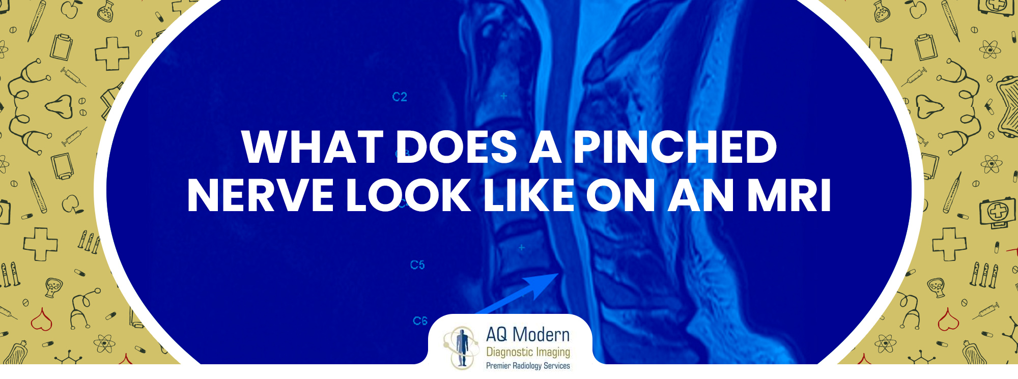
What Does a Pinched Nerve Look Like on an MRI? Unveiling the Images and Understanding Diagnosis
Have you ever experienced tingling, numbness, or shooting pain down your leg or arm? It could be due to pinching of nerves, a common condition caused by compression on a nerve. While symptoms can be telling, a definitive diagnosis often relies on imaging techniques like Magnetic Resonance Imaging (MRI). But how exactly does a pinched nerve show up on an MRI?
This article delves into the world of MRIs and pinched nerves, exploring what radiologists look for in these scans and how they interpret the findings. Furthermore, we’ll also address the limitations of MRIs and discuss other diagnostic tools. Finally, the article concludes with resources for high-quality imaging centers like NJ Imaging Center and its open MRI services in Edison, New Jersey.
Can MRIs Detect Nerve Damage?
MRIs are effective diagnostic instruments that use radio waves and strong magnetic fields to provide precise cross-sectional pictures of the body. Although nerves themselves aren’t directly visible on an MRI, this imaging technique can provide valuable insights into the condition. Let’s examine MRIs’ role in the diagnosis of nerve pain in more detail:
Our bodies depend on nerves to transfer messages between the brain, spinal cord, and different organs. That is why when nerve compression or damage happens, it leads to symptoms such as numbness, tingling, weakness, and pain. A pinched nerve is one of the most common causes of nerve pain.
What to Expect on an MRI for a Pinched Nerve
When you undergo an MRI (Magnetic Resonance Imaging) to assess a suspected pinched nerve, the procedure typically focuses on examining the spine—the most common location for nerve compression. Here’s what you can expect:
Preparation and Positioning:
- You will be placed on a table that glides inside the huge cylindrical MRI scanner.
- Technicians will use straps and coils to position you comfortably and ensure optimal image quality.
- In order to get clear images from the scan, you need to be still.
The MRI Experience:
- The MRI machine generates loud clicking noises throughout the scan. These noises are normal and part of the imaging process.
- Furthermore, the scan might take anywhere between 15 and 45 minutes to complete.
- To make the experience more pleasant, you may be provided with headphones playing calming music to mask the noise.
Addressing Claustrophobia:
- If you’re prone to claustrophobia (fear of enclosed spaces), consider open MRI scanners.
- Facilities like NJ Imaging Center in Edison offer open MRI options, which provide a more spacious and less confining environment.
Interpreting the Images: Signs of a Pinched Nerve on MRI
Radiologists and medical professionals trained in interpreting medical images analyze MRI scans. Here’s what they typically look for when evaluating a pinched nerve:
Disc Herniation: The discs in your spine serve as cushions between the vertebrae. A ruptured or bulging herniated disc may obstruct the nerves that are connected to the spinal canal. On an MRI, a herniated disc might appear displaced or misshapen, potentially narrowing the space around the nerve root.
Bone Spurs: These bony outgrowths on the vertebrae can also compress nerves. MRIs can reveal the location and size of bone spurs, helping doctors understand their role in nerve compression.
Spinal Stenosis: Additionally, pressure can be applied to the spinal cord and nerve roots due to a narrowing of the spinal canal caused by several conditions, including arthritis. MRIs can show a decrease in the normal space within the spinal canal, suggesting spinal stenosis.
Inflammation: While not always visible, an MRI might reveal signs of inflammation around the nerve, which can contribute to pinching.
Limitations of MRIs for Pinched Nerves
While MRIs are highly valuable for diagnosing pinched nerves, it’s important to understand their limitations:
Nerve Damage Visibility:
- MRIs are excellent at visualizing structural abnormalities, including nerve compression. However, they may not always detect subtle nerve damage or inflammation, particularly in the early stages.
- Mild nerve injuries or minor inflammation might not be apparent on MRI scans. Therefore, if a patient experiences symptoms suggestive of a pinched nerve but the MRI appears normal, further evaluation may be necessary.
Pain Source Accuracy:
- While an MRI can reveal anatomical details, it doesn’t always pinpoint the exact source of pain.
- Sometimes, a pinched nerve seen on an MRI may not be the sole cause of discomfort. Furthermore, other factors, such as muscle strain, joint issues, or inflammation, could contribute to the overall pain experience.
- Therefore, clinical correlation—combining MRI findings with the patient’s symptoms and physical examination—is crucial for accurate diagnosis and effective treatment.
Beyond the MRI: Additional Diagnostic Tools for Pinched Nerves
For a thorough assessment, an MRI may occasionally be supplemented by other diagnostic procedures:
X-rays: Though not ideal for visualizing soft tissues like nerves, knee X-rays can reveal bone abnormalities that might contribute to nerve compression.
Electromyography (EMG) and Nerve Conduction Studies (NCS): These tests measure electrical activity in muscles and nerves, helping assess nerve function and identify the location and severity of damage.
Treatment Options for Pinched Nerves
Treatment for pinched nerves depends on the severity and location of the compression. Common approaches include:
Rest and Activity Modification: Reducing strain on the affected area can promote healing and alleviate pain.
Medication: Anti-inflammatory medications and pain relievers can help manage inflammation and discomfort.
Physical Therapy: Physical therapy exercises can strengthen muscles, increase nerve function, and increase flexibility.
Injections: Steroid injections can relieve pain by reducing inflammation surrounding the pinched nerve.
Surgery: In extreme situations where conservative measures are ineffective, the nerve may need to be operated on to eliminate the source of its compression.
Conclusion: Finding Relief with Advanced Imaging and Expert Care
A pinched nerve can be a frustrating and painful condition. While an MRI is a valuable tool for identifying the underlying cause, it’s just one piece of the diagnostic puzzle. That is why consulting with a qualified healthcare professional who can interpret your MRI findings alongside other tests and your symptoms is crucial for an accurate diagnosis and effective treatment plan.
If you are looking for an open MRI in Edison, AQ Imaging is your reliable destination. At NJ Imaging Center, we use state-of-the-art facilities to ensure accurate results.

