For years, the constant advancement in medical imaging practices and procedures has caught the attention. These are widely beneficial in enhancing the diagnosis and medical treatments. A head MRI is also one of the painless and non-invasive techniques. It produces detailed images of the brain and brain stem.
For a head MRI, the radiologists use a magnetic field and radio waves. Open Mri for Head can reveal whether you have suffered any damage due to a stroke or a head injury. The MRI scans your brain using powerful magnets and computers. Also, it’s a bit different from a brain MRI. MRI images are more exact and crisper than other types of diagnostic imaging. Read on if you want to know more about the MRI procedure for the head and the purpose of MRI head scans. We have summed up all details to help you learn more about head MRI scans.
Why do you need a Head MRI?

A head MRI helps detect different brain conditions and diseases. It may be suggested by your consultant to discover signs such as:
- Dizziness
- Weakness
- Convulsions
- Mental or behavioral changes
- Eyesight problems
- Headaches for a long time
These symptoms could indicate a brain problem, which an MRI scan can identify.
Your doctor may command an MRI to inspect “portions” of your brain to decide what’s causing your symptoms. An MRI can assist your doctor in making an initial analysis of numerous health concerns.
A head MRI may be ordered so that your doctor can check for any of the following:
- Infections
- Stroke
- Tumors of the brain
- Multiple sclerosis (MS) is a disease that affects people
- Bleeding or hemorrhage in the brain
- Disorders of the eyes or the inner ear
- Disorders of the pituitary gland
- Clots in the blood
- Aneurysm, or a bulging of the blood arteries in the brain
- Anomalies in development
- Injuries to the spinal cord
- Cysts
- Swelling
- Inflammation
- Hormonal imbalances
- An accumulation of unsolidified in the brain is recognized as hydrocephalus.
MRI Procedure for Head

Maintaining complete stillness throughout the exam is critical to capturing the finest photos. Either orally or through an IV line, sedation may be required for children who have difficulties staying still. Adults who feel claustrophobic may benefit from sedation.
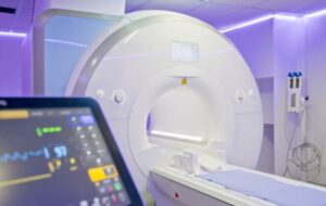
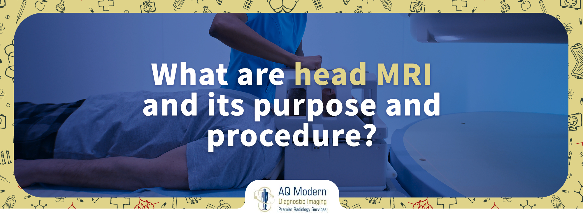
You’ll be positioned on a table that glides inside the MRI machine. Then, a vast magnet moves the table in the shape of a tube. A plastic coil may be fitted around your head.
A technician will take many photographs of your brain after the table is pushed into the machine, which will take a few minutes.
The machine will have a microphone so you may converse with the staff. It usually takes 30 to 60 minutes to complete the test. An IV may be used to administer a contrast solution. Commonly, Gadolinium is used. It helps the head MRI be more explicit, reflecting certain brain regions. It mainly focuses on the blood arteries.
During the operation, the MRI scanner will generate loud pounding noises. You may be provided with earplugs to block out the sounds from the MRI scanner. A head MRI does not pose any hazards.
A contrast solution may cause an allergic reaction in a small percentage of people. If your kidney function has deteriorated, inform the doctor or radiologist. As in this case, the contrast solution may not be safe.
A radiographer trained in imaging studies operates the MRI scanner. They are responsible for keeping the computer away from the magnetic field generated by the scanner. They control it from another room.
During the scan, you’ll be able to converse with the radiographer over an intercom, and they’ll be able to observe you on a monitor.
What Does a Head MRI Display?

MRI can be used to scrutinize almost every part of the body. MRI images of soft tissues, such as the brain, are extremely detailed. Because air and hard bone do not produce an MRI signal, they appear black.
The amount of fat and water present in each tissue and the machine settings utilized for the scan affect the intensity of bone marrow, spinal fluid, blood, and soft tissues, ranging from black to white.
Brain MRI scans may also be used to provide more detailed information about head procedures. Similarly, after surgery, they may be required to confirm that the healing process is going smoothly. Accidents and head injuries also prompt doctors to order scans of the brain to look for injuries, bleeding, and edema.
Most people are concerned about feeling claustrophobic. It happens when a person is placed in a small space, such as an MRI tube, and has anxiety. Although mirrors may be beneficial, some people may require medicine to help them relax.
Final Thoughts
The bottom line is that head MRI is safe. There are no known health risks linked with the magnetic field or the open MRI for a head scan. The overall MRI procedure for the head is painless and takes 30 to 40 minutes. A professional and trained radiologist will be present throughout the procedure to guide and complete the MRI head scans.
If you have been suggested to have an open MRI for a head scan, don’t worry about it. You have to look for a reputable imaging center near you. They will carry out the process smoothly.

