lighting the MRI Hyperintensity
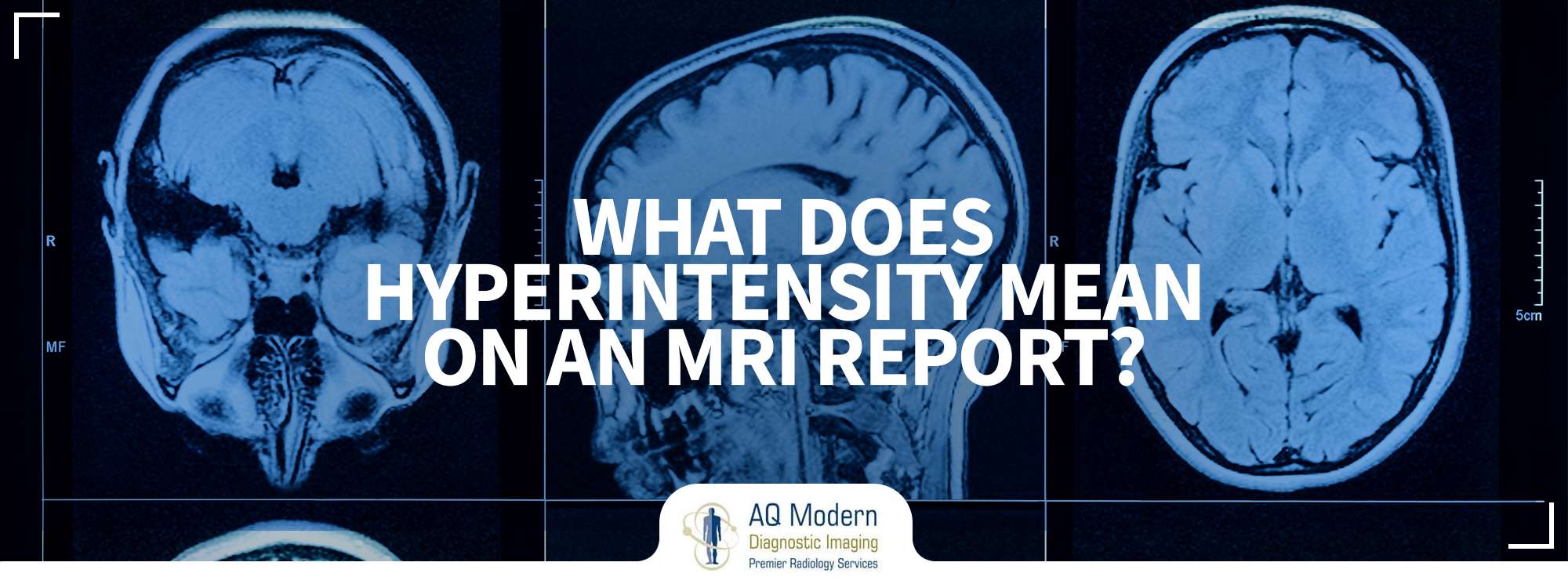
The term MRI hyperintensity defines how components of the scan look. Most MRI reports are black and white with shades of gray. However, the hyperintensity area appears a little lighter comparatively. On the contrary, hypo intensity would be blacker in color.
The MRI hyperintensity reflects the existence of lesions in the brain. The MRI imaging presents a range of sequences. These also involve different imaging patterns that highlight the different kinds of tissues. As a result, it makes it easier to detect abnormalities.
All over the world, an MRI scan is a common procedure for medical imaging. Since its invention, researchers and health practitioners are constantly refining MRI imaging techniques. It has significantly revolutionized medicine. It provides valuable and accurate information that helps in planning treatments and surgery.
Magnetic Resonance Imaging involves the use of a resilient magnetic field and radio waves. It produces images of the structures and tissues within the body. Overall, it’s a non-invasive and painless method that provides a detailed and cross-sectional illustration of the internal organs.
MRI scan is different from other diagnostic imaging techniques. Radiologists are responsible for imaging and developing MRI reports that help assesses and evaluate the health condition. It is an accurate method of detecting and confirming the diagnosis. Largely it defines the brain composition and weighs the reliability of the spinal cord. It also assesses the structure of the heart and aorta.
Do you Know What Hyperintensity Means on an MRI Report?
An MRI scan is one of the most refined imaging processes. As a result, it has become increasingly valuable in diagnosing health issues. It provides excellent visuals of soft tissue and allows the diagnosis of the following:
- Strokes
- Bleeds
- Tumors
Doctors measure hyperintensity by evaluating the imaging reports. It helps in detecting different mental disorders. For example, when MRI hyperintensity is 2.5 to 3 times, it indicates major depressive disorder or bipolar disorder.
The T2 MRI hyperintensity is often a sign of demyelinating illnesses. . MRI hyperintensity on a T2 sequence reflects the difference in the brain tissue at one part of the brain compared to the rest. When MRI hyperintensity is bright, clinical help becomes critical.
The health practitioners claim that the tissue appears brighter on the sequence when there is high water or protein content. Therefore, it is identified as MRI hyperintensity.
In medicine, MRI hyperintensity is available in three forms according to its location on the brain. These include:
- Profound white matter intensities.
- Periventricular white matter hyperintensities
- Subcortical hyperintensities.
also read: Is Mri Save Or Not
The MRI hyperintensity is an autoimmune illness. It affects the brain of humans and is more prevalent in older people. It has become common around the world. By highlighting the importance of managing the quality of MRI scans and images. As technology advances, radiologists are bringing new MRI techniques and machines to the market. In the United States, you can find a network of imaging centers that facilitate patients. They offer high-quality diagnostic services that enable the treatments.
However, it also exists in young and middle-aged people who have a history of other medical issues. In addition, practitioners associate it with cerebrovascular disorders and other similar risks. Therefore, the doctors focus on neurological evaluation when assessing the MRI reports providing the diagnosis accordingly.
What are the Reasons for MRI Hyperintensity?
It’s not easy for common people to understand the neuropathology of MRI hyperintensity. However, it is commonly associated with the following vascular risk factors:
- Smoking
- Hypertension
- Diabetes
The white MRI hyperintensity is often a reflection of small vessel disease. Some of the associated neuro-pathological issues are:
- Ischemic damage
- Cerebral atrophy
- Axonal loss
- Hypoperfusion
- Hypoxia
- Reduced glial density
In this case, it’s essential to understand the clinical significance of MRI hyperintensities. The white matter MRI hyperintensities help in assessing and confirming the existence of the vascular disease. In such cases, high blood pressure and age are key risk factors.
Weakened flexibility and reduced cognitive function are often a result of white matter MRI hyperintensity.
On the other hand, it has a sturdy impression on memory and executive running. However, the level of impact relies on the severity and localization of the MRI hyperintensity.
The health practitioners also state that MRI hyperintensity is also associated with a decline in cognitive behavior. For example, it affects the handing out speed and executive functions.
According to health practitioners, there is a strong connection between death and MRI hyperintensity. The risk is high in people with a history of stroke and depression. The presence of hyperintensity leads to an increased risk of dementia, mortality, and stroke. It is also linked with constant and resistant depression.
The MRI scan helps doctors in examining the health of the brain. They can screen the risk factors, making it easier to opt for proactive measures that can help treat an illness.
Open and Closed MRI and MRI Hyperintensity
Suppose you are having a medical issue, and your physician recommends an MRI. You don’t need to panic as most laboratories have advanced wide-bore MRI and open MRI machines. It helps in accurately diagnosing and assessing the diseases.
On the other hand, the wide-bore MRI scanner also provides accurate and high-quality images. It’s beneficial in case patients are claustrophobic. The wide space makes it easier to conduct brain MRI and other body parts as required.
The open MRI involves an open machine that uses magnets to take inside images from all four sides.
As compared to ultrasound and CT scans, MRI has more advantages. It provides a more clear and visible image of the tissues. The MRI hyperintensity is the white spots that highlight the problematic regions in the brain. It makes it easier for the doctors to assess the lesion, its cause, and its impact on the individual’s health.
The MRI hyperintensity is a common imaging feature in T2 MRI imaging reports. It also indicates the effects on the spinal cord. Both the wide bore and open MRI scan methods help radiologists in narrowing the diagnosis. The doctors also integrate patients’ medical history and evaluate the laboratory test results accordingly for clarification and authentic assessment.
Final Thoughts
The MRI hyperintensity reflects the existence of lesions on the brain of the individual. It is a common imaging characteristic available in magnetic resonance imaging reports. Using MRI scans as a diagnostic approach helps in managing effective clinical evaluation. It also acts as a practical framework that allows the radiologists to plan the overall treatment.
When examining the MRI scan, doctors and radiologists look for the MRI hyperintensity. It indicates the lesions, their volume, and their frequency. The assessment of the MRI hyperintensity lesions assists in diagnosing neurological disorders and other psychiatric illnesses.
Overall, the MRI scans are highly beneficial in detecting health disorders, allowing proactive designing of the treatment plans. Therefore, healthcare providers need to interpret the imaging reports and provide their patients with relevant information to help them understand their health conditions.

